Palmar Aponeurosis of Hand
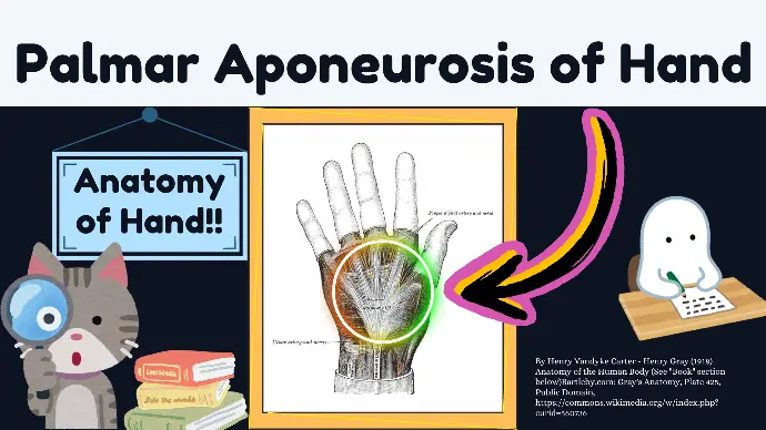
The palmar aponeurosis is a thick, fibrous structure in the palm of the hand that plays a crucial role in hand function and mechanics.
It is the central part of the deep fascia of the palm.

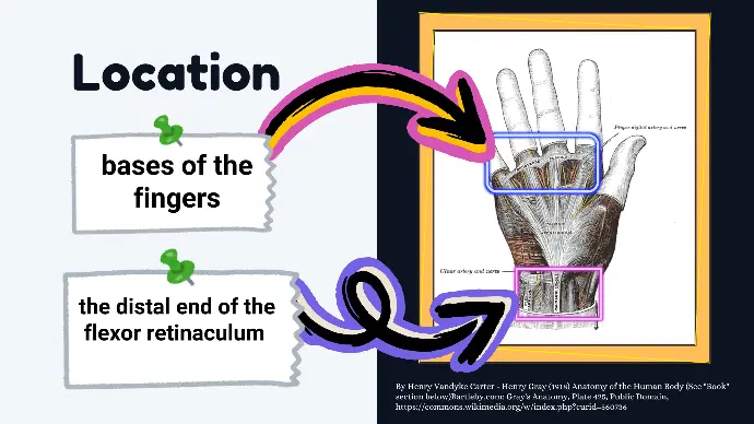
Location of Palmar Aponeurosis
The palmar aponeurosis is centrally located in the palm, extending from the distal end of the flexor retinaculum to the bases of the fingers. Its central position allows it to connect and support various structures within the hand.

Structure of Palmar Aponeurosis
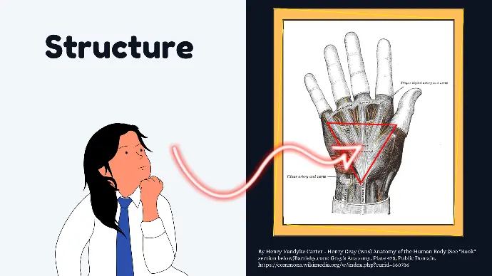
It is triangular in shape, with:
- Apex: Directed proximally towards the wrist, where it is continuous with the flexor retinaculum and the tendon of the palmaris longus muscle.
- Base: Directed distally towards the fingers, where it divides into superficial and deep layers, forming four slips that extend into the fingers.

Anatomy of Palmar Aponeurosis
- Proximal Attachments: The apex is continuous with the flexor retinaculum and the palmaris longus tendon, anchoring the aponeurosis to the wrist.
- Distal Attachments: The base is divided into superficial and deep layers. The deep layer forms four digital slips that extend towards the medial four digits, blending with the fibrous digital sheaths.
- Distal Extensions: These four digital slips extend into the fingers, providing stability and blending with the fibrous sheaths surrounding the flexor tendons.
Layers of Palmar Aponeurosis
- Superficial Layer: This layer is attached to the dermis of the skin, providing a connection between the skin and the underlying structures.
- Deep Layer: Composed of longitudinally oriented fibrous bands, this layer extends distally to form the four digital slips that attach to the fibrous sheaths of the fingers.
Anatomy of Palmar Aponeurosis
in Relation to Other Structures
- Flexor Retinaculum: The proximal end of the palmar aponeurosis is continuous with the flexor retinaculum, which forms the roof of the carpal tunnel.
- Flexor Tendons: The aponeurosis overlies the flexor tendons, providing them with a protective covering and aiding in their function.
- Nerves and Vessels: The aponeurosis protects the median and ulnar nerves, as well as the superficial and deep palmar arches and their branches.
Clinical Pathology
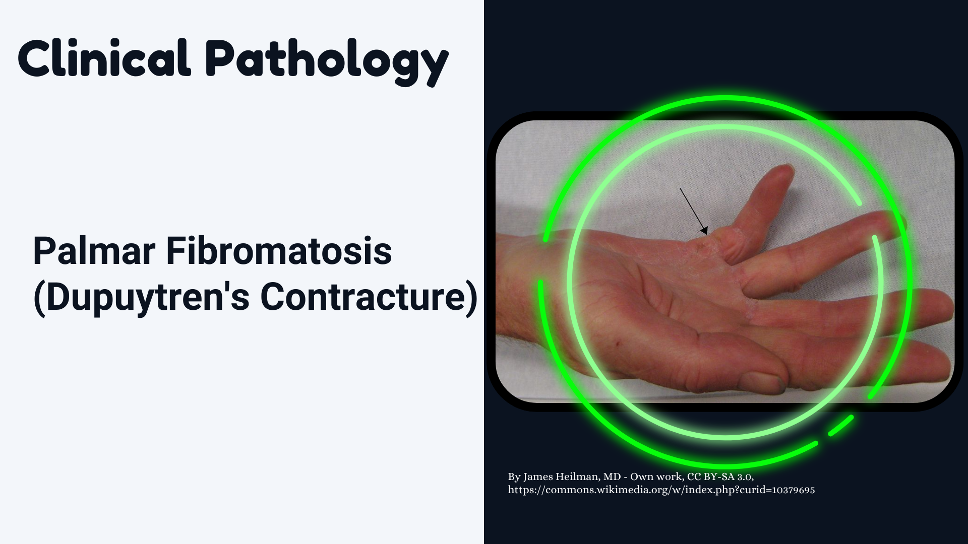
Palmar Fibromatosis
(also known as Dupuytren's Contracture)

It is a pathological condition where the palmar aponeurosis becomes thickened and contracted, leading to flexion deformities of the fingers, causing them to bend towards the palm and limiting hand function. The condition often progresses over time and may require medical intervention, such as surgery, to restore normal hand function.