The Shoulder Joint
The shoulder joint is a “Ball and Socket” Type of Synovial Joint.
Therefore, it has a wide range of movements.
There is no joint in the body, that has more mobility than the shoulder joint.
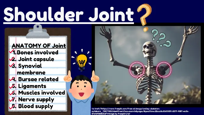
Anatomy of the Shoulder Joint

The shoulder joint, also known as the glenohumeral joint, is one of the most complex and mobile joints in the human body.
We will discuss the following points in the anatomy of shoulder joint:
- Bones involved
- Joint capsule
- Synovial membrane
- Bursae related to joint
- Ligaments
- Muscles involved
- Nerve supply
- Blood supply
1. Bones Involved
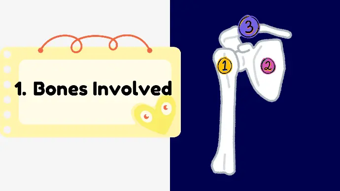
Three bones are involved in Shoulder Joint.
- Humerus: The proximal end of the humerus forms the ball of the ball and socket joint.
- Scapula: The glenoid cavity of the scapula forms the socket of the ball and socket joint.
- Clavicle: Connects the shoulder to the sternum, acting as a strut to stabilize the shoulder.
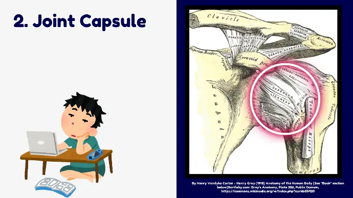
2. Joint Capsule
The joint capsule is a fibrous envelope that encloses the structures of the shoulder joint, providing stability and flexibility.
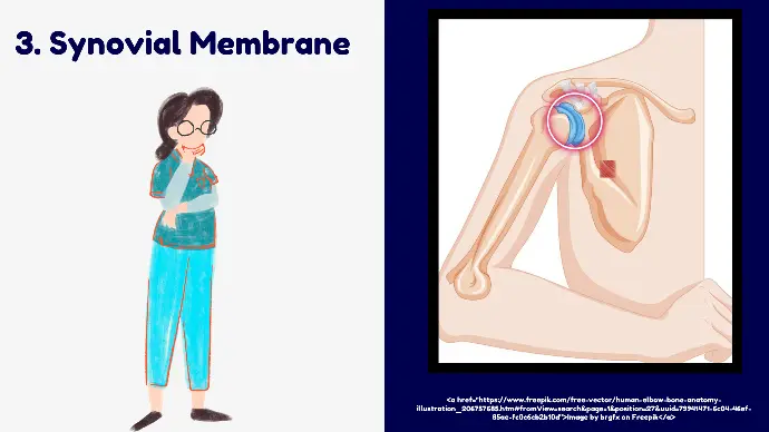
3.Synovial Membrane
The inner lining of the joint capsule secretes synovial fluid, which lubricates the joint.
4.Bursae related to Joint
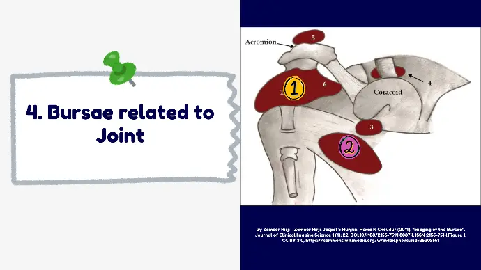
These are sacs filled with Synovial fluid, that surrounds the shoulder joint at specific locations.
There are 3 Bursae related to Shoulder Joint.
- Subacromial Bursa (it is also known as Subdeltoid Bursa).
- Subscapularis Bursa
- Infraspinatus Bursa
5.Ligaments related to Joint
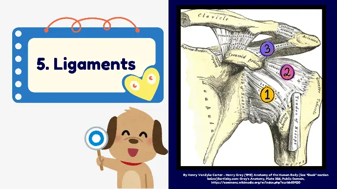
- Glenohumeral Ligaments:
these are Superior, Middle, and Inferior Glenohumeral Ligaments. They Strengthen the anterior aspect of the joint capsule.
- Coracohumeral Ligament:
Extends from the coracoid process of the scapula to the greater tubercle of the humerus. It gives strength to the capsule.
- Coracoacromial Ligament:
Forms the coracoacromial arch, which prevents superior displacement of the humeral head.
- Transverse Humeral Ligament:
It bridges the bicipital groove of humerus. Holds the long head of the biceps tendon in place within the bicipital groove.
6.Muscles Involved in Joint
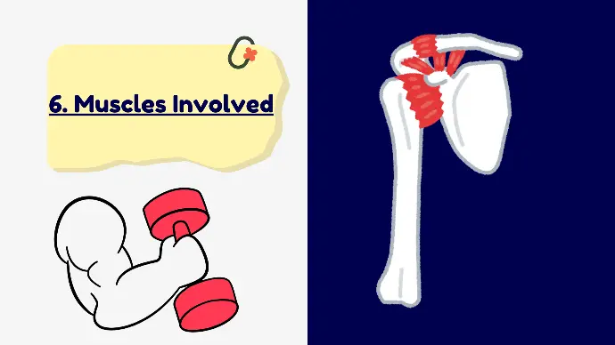
It has 2 categories of Muscles involved:
- Rotator Cuff Muscles
- Other Muscles
1. Rotator Cuff Muscles
These are:
- Supraspinatus
- Infraspinatus
- Teres Minor
- Subscapularis
2. Other Muscles:
These are
- Deltoid
- Pectoralis Major
- Latissimus Dorsi
- Teres Major
- Biceps Brachii
- Triceps Brachii
7. Nerve Supply of Shoulder Joint
Four Nerves are involved in Shoulder Joint.
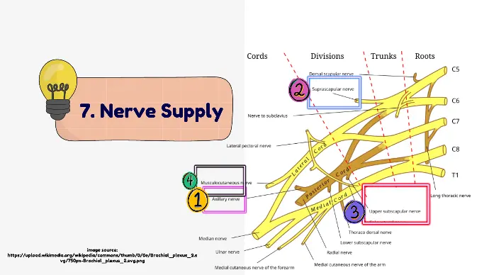
- Axillary Nerve: Innervates the deltoid and teres minor muscles.
- Suprascapular Nerve: Innervates the supraspinatus and infraspinatus muscles.
- Subscapular Nerves: Innervate the subscapularis and teres major muscles.
- Musculocutaneous Nerve: Innervates the biceps brachii and other flexor muscles of the arm.
8. Blood Supply of Shoulder Joint
Following Arteries supply the Shoulder Joint.
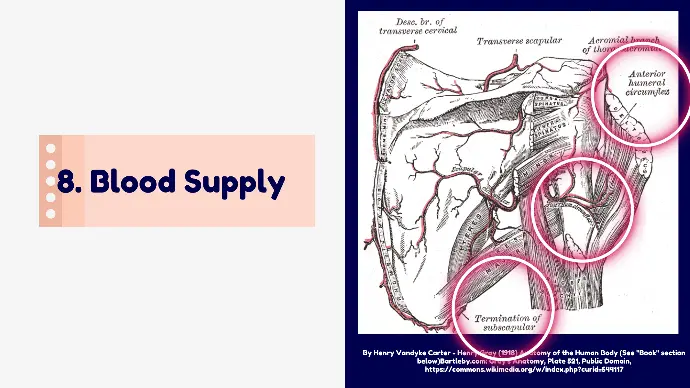
- Anterior and Posterior Circumflex Humeral Arteries: Branches of the axillary artery.
- Suprascapular Artery: Branch of the thyrocervical trunk.
- Subscapular Artery: Branch of the axillary artery.
Clinical Significance
Following are the Common Pathologies of Shoulder Joint.
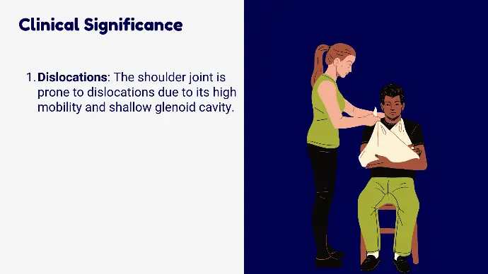
1. Dislocations of Shoulder joint:
The shoulder joint is prone to dislocations due to its high mobility and shallow glenoid cavity.
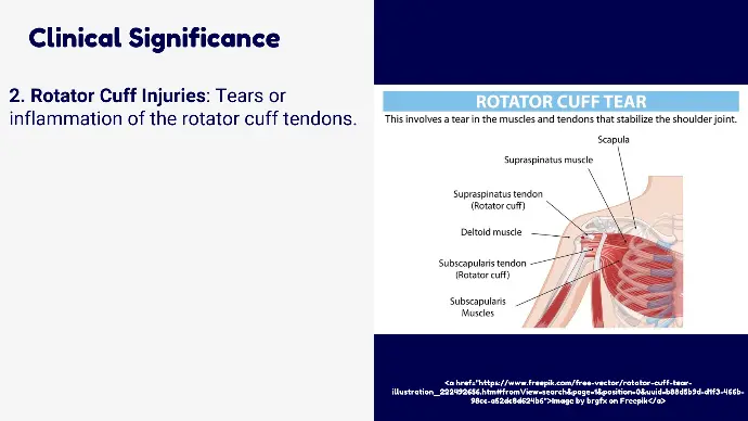
2. Rotator Cuff Injuries:
Tears or inflammation of the rotator cuff tendons.
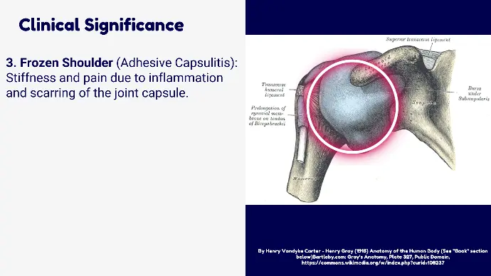
3. Frozen Shoulder
(Adhesive Capsulitis):
Stiffness and pain due to inflammation and scarring of the joint capsule.
4. Arthritis of Shoulder joint:
Degenerative changes in the joint leading to pain and reduced mobility.
